Dental cavities, a prevalent dental issue worldwide, affect millions of people annually. These cavities result from decay and bacterial activity that gradually breaks down the tooth’s enamel, forming holes or cavities. To rectify this, dental fillings are necessary. Ignoring these cavities can lead to severe dental problems and cause considerable pain and discomfort in the process. As cavities aren’t always visible to the naked eye, dentists advise getting annual X-rays. These X-rays provide additional information beyond what a clinical oral examination can reveal. They help detect proximal dental caries (cavities between teeth) and early carious lesions (tiny cavities) that may be too small to detect otherwise. Early detection allows for cavity reversal, preventing the need for costly dental procedures.
Table of Contents
What Do Cavities Typically Appear Like on an X-Ray?
Cavities visible on X-rays differ from those in the mouth due to the black-and-white nature of X-rays. When examining an X-ray, keep an eye out for the following:
- Cavities: Cavities on X-rays are known as radiolucencies. These dark areas on an X-ray indicate the presence of hollow spaces, which are essentially cavities within the teeth.
- Healthy Teeth: Healthy tooth structures appear more radiopaque or denser. This means they appear brighter on an X-ray.
- Fillings/Dental Work: Fillings appear even more radiopaque than the surrounding tooth structure and often have an extremely white appearance (although this depends on the material used). Specifically designed to differ significantly from regular teeth, fillings help dentists distinguish between healthy tooth structures and past dental work.
X-Rays of Teeth Without Cavities
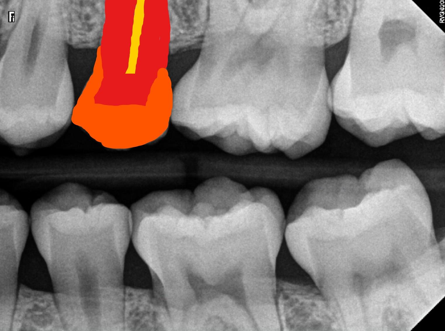
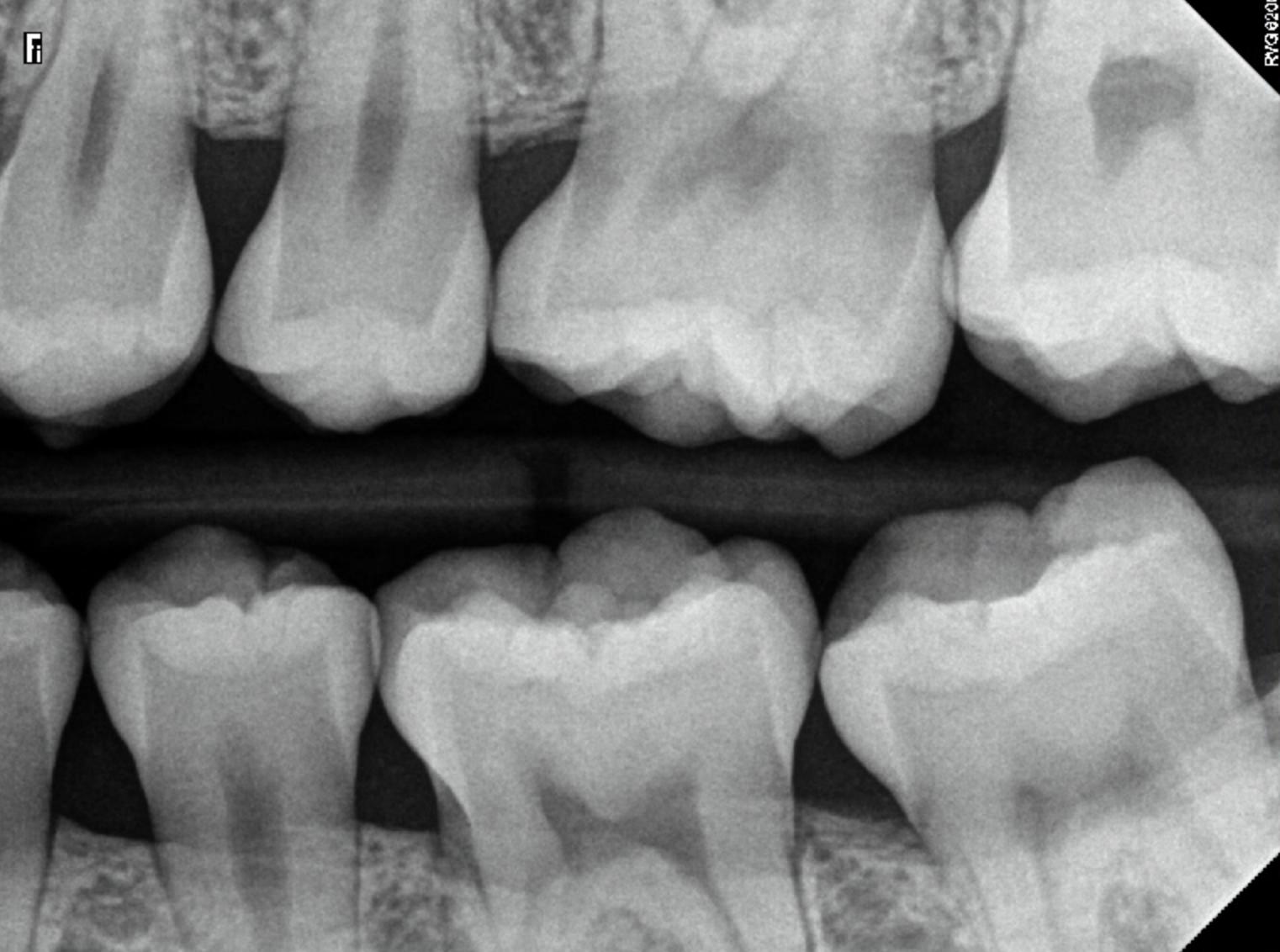
Below is an image depicting teeth without any cavities, providing a reference for comparison. The left image shows teeth with no fillings, representing a fairly healthy tooth’s appearance on an X-ray. The outermost layer, known as the enamel, appears very bright and is the most radiopaque. The middle layer, known as dentin, also appears bright but less so than the enamel. The innermost layer of the tooth, called the pulp, appears the darkest on an X-ray. In the image, we have highlighted these areas with colors for better understanding: yellow for the pulp, red for dentin, and orange for enamel.
X-Ray of Teeth Under Observation (Mild Cavity)
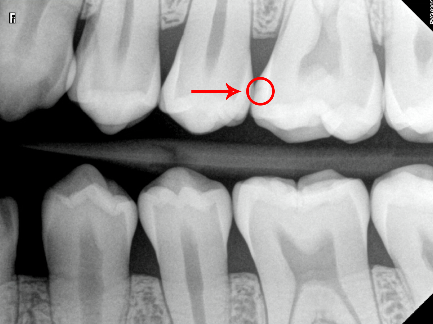
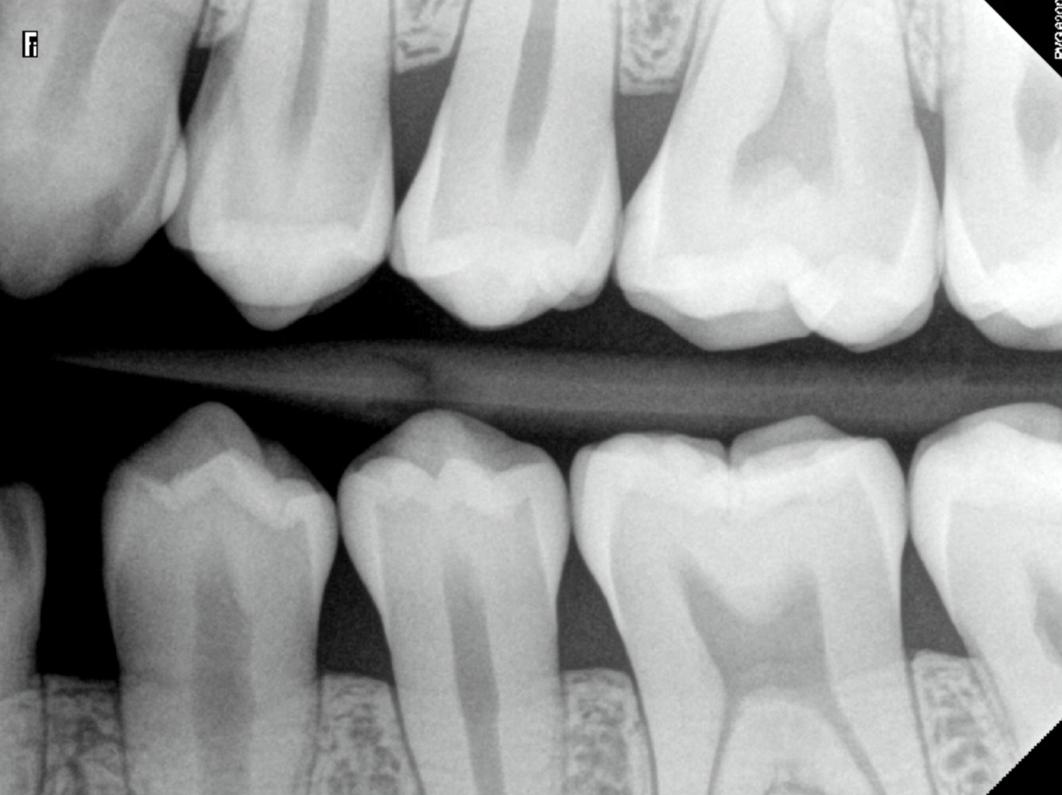
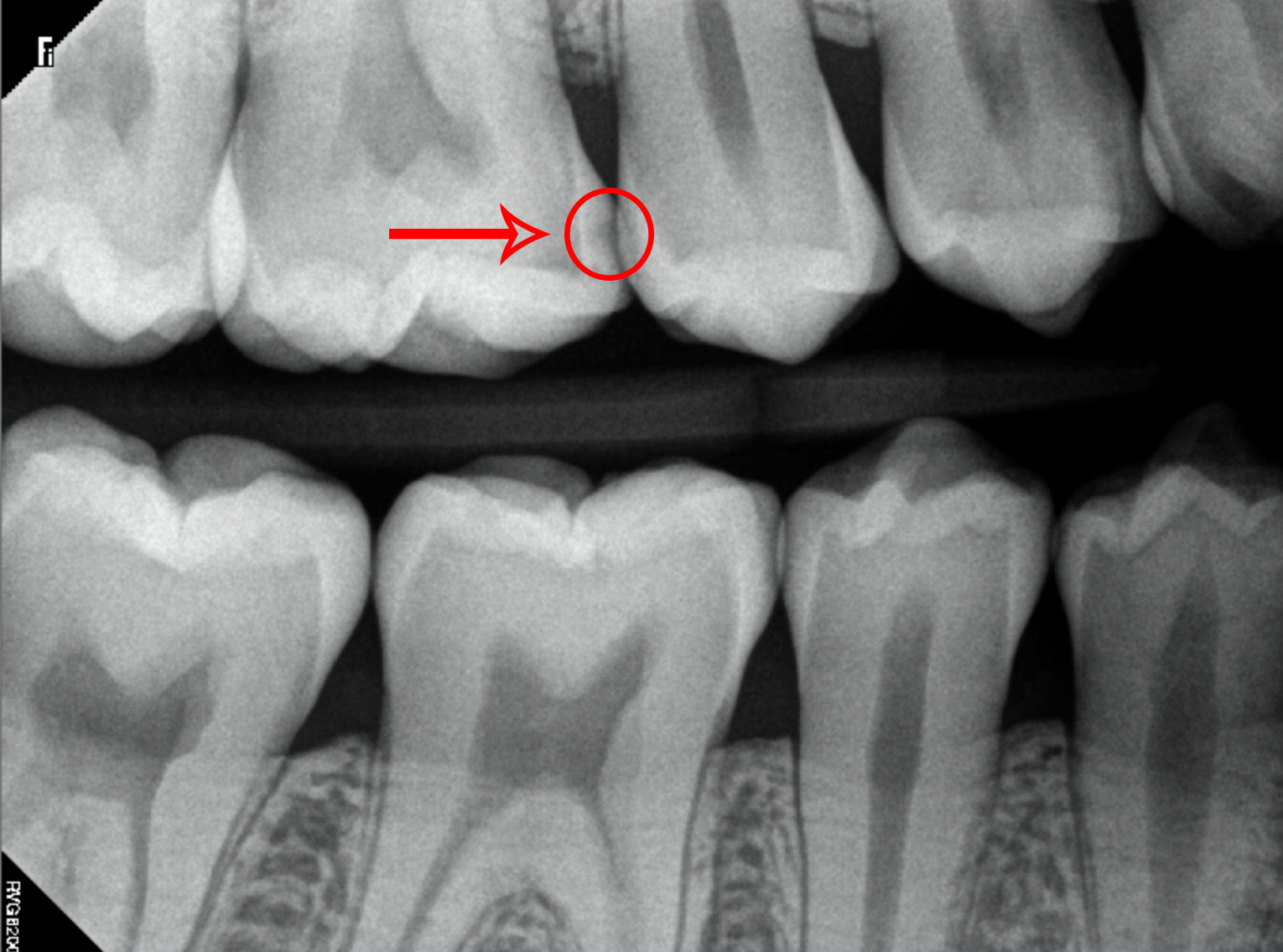
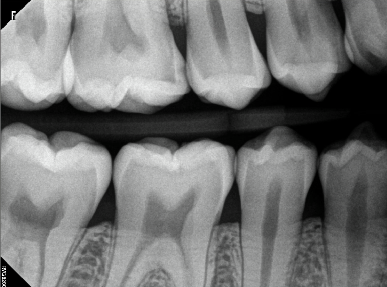
Teeth under observation have cavities in their earliest stages, having not yet penetrated the enamel surface. These cavities may be challenging to spot on an X-ray but typically appear as tiny and faint radiolucencies. With appropriate treatment, these cavities can be reversed, preventing further damage. In the image below, we focus on the upper left first molar (tooth number 14 for the American tooth numbering system or 26 for the Canadian tooth numbering system). It shows a tiny radiolucency just beginning to form on the mesial surface, which is the part closest to the center of the mouth, located between the teeth.
X-Rays of Teeth With Moderate Cavities
As cavities progress and become more severe, they become more visible on X-rays. These cavities extend deeper into the tooth, past the enamel and into the dentin. They typically appear as darker areas on X-rays. Some cavities may even exhibit irregularly shaped “holes” compared to teeth without cavities. Dental fillings are necessary to restore these types of cavities properly. In the image below, we see the upper right first molar (tooth number 3 for Americans or 16 for Canadians) with a significantly sized cavity visible on the X-ray. This cavity is also located on the mesial surface and is more easily detected because it has penetrated beyond the enamel layer.
In the same image, a child exhibits multiple cavities. Of particular interest is the cavity on their upper right second molar (tooth A for Americans or 55 for Canadians). This cavity appears as a darker area compared to the other teeth and is situated on the occlusal surface of the tooth, which is the part you bite down on.
X-Rays of Teeth With Severe Cavities
Untreated cavities will continue to spread deeper into the tooth, eventually consuming the entire tooth structure, including the enamel, dentin, and pulp. On X-rays, these severe cavities appear as large dark areas. They cause intense pain and necessitate root canal treatment or extraction. In the image below, the patient exhibits cavities on all of their teeth. We focus on the upper right first molar (tooth number 3 for Americans or 16 for Canadians), which shows a severely large cavity on the distal surface. This cavity is easily noticeable as it has eroded a significant portion of the tooth structure.
Returning to the previous image featuring the child with multiple cavities, there is also a cavity that has reached the pulp. We now pay attention to the cavity on their lower right second molar (tooth T for Americans or 85 for Canadians). This occlusal cavity is much more severe, appearing as a large dark area on the X-ray, with the majority of the tooth structure eroded away. In children, a baby root canal or extraction is typically recommended.
Conclusion
In conclusion, cavities can vary in severity, and their appearance on X-rays reflects this. Beginning cavities may appear as tiny and faint radiolucencies, while severe cavities exhibit a much darker appearance. By understanding how cavities appear on X-rays, dentists can detect and treat them at early stages, preventing more severe damage in the future.
For more information about dental health, visit 5 WS.

